

OVERVIEW
The term hypertensive retinopathy describes the damage caused to the retinal blood vessels due to long-term high blood pressure. The retina is the light-sensitive layer at the back of the eye that is responsible for vision. With damage to the retina, hypertensive retinopathy can severely damage vision causing even loss of sight.
Ishwar Eye Centre, a leading Hypertensive Retinopathy Hospital in Rohtak, offers specialized care and early intervention for such retinal damage.
Vision is determined by the ability of the retina to receive the light signals and convert them into electrochemical signals that are transmitted to the brain via the optic nerve. The retina is a layer of highly sensitive cells that are rich in blood supply and nerves.
Retina and blood pressure
Retinal blood vessel changes depends on two major factors –
- level of the blood pressure
- state of the arterioles or the small blood vessels that supply the retinal cells
Ishwar Eye Centre, being a prominent Hypertensive Retinopathy Hospital in Rohtak, continuously monitors these factors using state-of-the-art diagnostic tools.
Causes
Due to rise of blood pressure there is spasm or narrowing of the tiny blood vessels in the retina. This may be seen in younger patients with healthy arteries as well.
The condition of arteries change with age and along with long term high blood pressure. This is called arteriolar sclerosis. This condition literally means hardening of the walls of the arteries and makes their walls less elastic to the blood flow. Hardened walls of the small arteries also make them prone to rupture in case of a high blood pressure flow through them.
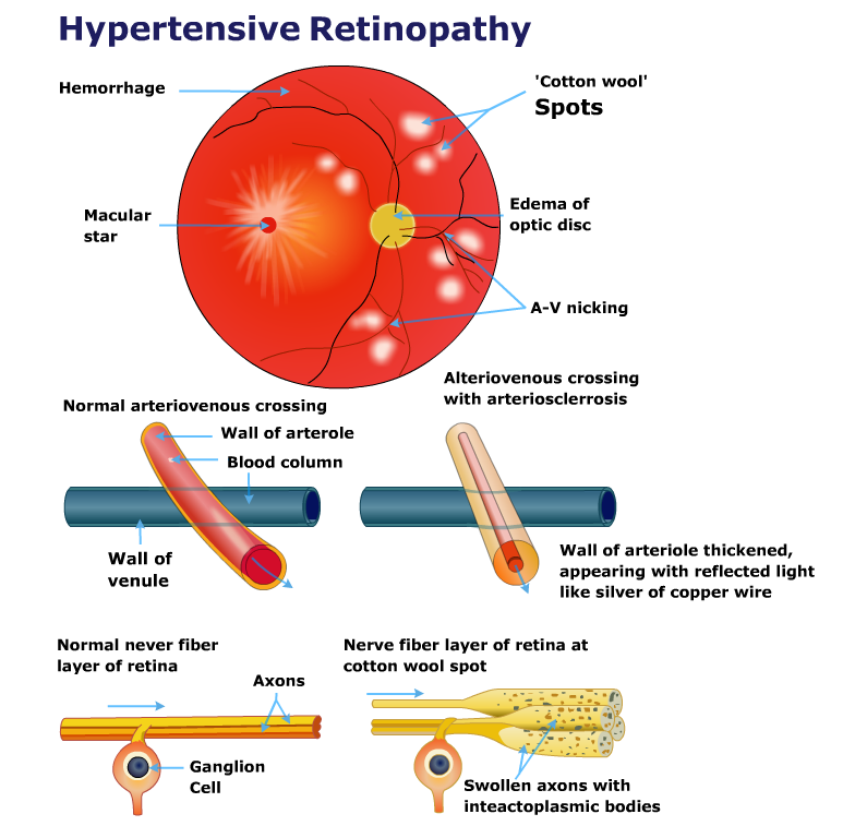
In elderly patients as well as those with essential hypertension some changes in the arterioles are seen. These are called silver wiring and AV nipping (atrioventricular nipping). These may be small bleeding spots where the arterioles have ruptured in the retina. This is called retinal hemorrhage. There are other features called cotton wool spots and flame haemorrhages along with swelling of the optic disc that are commonly seen in young patients with malignant hypertension.
Patients dealing with these conditions are advised to consult a dedicated Hypertensive Retinopathy Hospital in Rohtak like Ishwar Eye Centre for early diagnosis and prevention of complications.
Symptoms
Hypertensive retinopathy is a slowly progressive condition. Initial damage to vision thus may not produce any symptoms. Symptoms may appear late in the disease process, usually when much of the vision is lost. Some of the symptoms include –
- Blurred vision
- Double vision or seeing double of everything
- Dim vision or difficulty in seeing in poor light
- Headaches due to difficulty in focussing
- Floaters in vision
- Flashes of light in vision
- Sudden vision loss. This is a medical emergency when suddenly there may be a complete black out of vision
If you’re experiencing any of these signs, visit Ishwar Eye Centre, the top-rated Hypertensive Retinopathy Hospital in Rohtak, for expert ophthalmic evaluation.
Diagnosis
A detailed history of duration and severity of hypertension is obtained. This condition is diagnosed usually by examining the retina using an ophthalmoscope.
For examining the retina and taking its digital photographs, drops are given in the eyes to dilate the pupils as wide as possible. Then, an ophthalmoscope is used to examine the retina. The blood vessels are visible on the photograph. Features of narrowing of blood vessels, leaking fluid and haemorrhages or bleeding spots are visible. The degree of retina damage (retinopathy) is graded on a scale of 1 to 4.
Yet another test is called flurescin angiography. Here a special dye is injected into a vein in the arm. The dye travels to all blood vessels including those of the retina. Using special camera, pictures of the retina are taken. The dye lights up in the images showing the blood vessel abnormalities in the retina.
Such advanced diagnostic methods are routinely offered at Ishwar Eye Centre, your trusted Hypertensive Retinopathy Hospital in Rohtak, to help detect the condition at its earliest stage.
Complication
According to present norms there are basically two groups of hypertensive retinopathy –
- Compensated hypertensive retinopathy – This has further two grades grade 1 and 2. In this type there are increased tortuosity of the retinal arteries, copper or silver wiring, AV nipping etc.
- Accelerated hypertensive retinopathy – In this type the retina shows cotton wool spots, flame haemorrhages, and hard exudates around the macula (or macular star), edema or swelling of the optic disc along with impairment of vision. This may lead to complications such as central and branch, retinal vein and/or artery occlusion (CRVO or CRAO), ischemic optic neuropathy and Vitreous haemorrhage.
- Loss of vision
- Central and branch, retinal vein and/or artery occlusion (CRVO or CRAO)
- Ischemic optic neuropathy or damage to the optic nerve
- Vitreous haemorrhage or bleeding in the vitreous humour
To prevent such serious complications, early treatment at a reputed Hypertensive Retinopathy Hospital in Rohtak like Ishwar Eye Centre is crucial.